

Data acquisition system for Correlative X-ray Imaging
- Type:Master Thesis
- Supervisor:
- Field of Study:
Physics, mechanical/electrical/chemical/ physical engineering and related areas
Job description
At the Institute of Microstructure Technology, the CORREL infrastructure for correlative characterization is developed. It combines X-ray imaging and nuclear magnetic resonance techniques in a single device. This unique instrument will expand imaging capabilities for biomedical and materials science applications.
The standard X-ray imaging technique relies mainly on absorption contrast, where the difference in the absorption coefficient of the constituents is linearly related to the obtained signal. Therefore, only highly absorbing structures, like bones surrounded by muscles or metals surrounded by less dense materials such as polymers, provide a good imaging contrast. The contrast tends to be very poor for weakly absorbing materials, e.g., to study soft tissues or polymer blends.
The evolution of innovative methods, like phase-sensitive techniques, led to a significant improvement in image quality, thus overcoming the limitations of conventional X-ray imaging.
This Master's thesis will be performed in the CORREL laboratory, focusing on establishing the basic radiographic configuration consisting of an X-ray source and an X-ray detector. This setup will be the foundation for testing objects like optical components and developing new phase-sensitive approaches to combine in a further step with NRM.
Single-shot X-ray Imaging (SSI) is an example of the methods that will be explored after implementing the basic radiographic system. The SSI configuration is obtained by adding an optical element called a Hartmann mask to the basic radiographic setup (Fig. 1). The SSI makes it possible to obtain from a single image simultaneously absorption, differential-phase, and scattering contrasts.
The implementation of phase-sensitive imaging within the X-ray-NMR scanner will offer spectral along with enhanced imaging contrast. It will open opportunities for fast measurements, like monitoring dynamical processes and performing low dose imaging.
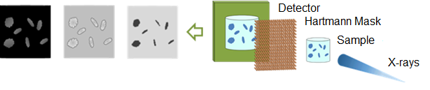
Fig. 1 Simplified depiction of the single-shot imaging setup. The setup shows the X-ray rays from the source, the sample to be characterized, a Hartmann mask as the main optical element, and the X-ray detector.
The aims of the master thesis are to
- Install the Xineos 2329 detector in the basic radiographic configuration, consisting of an X-ray source, test object, and detector;
- Communicate the detector with the computer;
- Develop the data acquisition (DAQ) software;
- Perform preliminary measurements and necessary experimental adjustments;
- Acquire and plot the source spot shape and the spot stability over 24 h.
The student will have the opportunity to acquire fundamental knowledge in X-ray imaging, while contributing to a cutting-edge research. Will have good supervision, be part of a highly motivated and friendly CORREL team. At the IMT, you have the opportunity to get to know several laboratories, including a cleanroom, and visit some installations at the Karlsruhe Research Accelerator (KARA).
Starting date: by appointment
Contract duration: 9 - 12 months
Qualification:
- Good academic record (marks);
- Strong interest in hardware/software for DAQ;
- Interested in lab work, engaged in research within a multidisciplinary environment;
- Enthusiastic, team player, innovative;
- Knowledge in lab instrumentation/imaging is desirable.
Technical Contact
Dr. Danays Kunka
Karlsruhe Institute of Technology
Institute of Microstructure Technology
P.O. Box 3640
76021 Karlsruhe
phone: +49 721 608-22193
e-mail: danays kunka ∂does-not-exist.kit edu