

Applied X-ray Imaging Systems (XIS)
Dr. Arndt Last [Contact]
Welcome
Our group develops X-ray optics and X-ray imaging techniques for use in materials science, medicine and research.
Our research contributes to the research program 3: "Materials Systems Engineering" (MSE), Topic 2: "Optics and Photonics: Materials, Devices and Systems" (Project 43.32.03, Refractive X-ray Optics), in the research field „Information“ as defined by the Helmholtz Association (link to MSE website). We contribute to the improvement of analytical and imaging techniques to answer the scientific and societal questions of our time.

Subject areas
Our research focuses on X-ray lenses made of polymers for beam shaping and for X-ray microscopy as well as X-ray gratings for phase-contrast imaging. The optics require the production of microstructures with a very high aspect ratio and extremely smooth surfaces, which is possible using X-ray lithography, for example.
Polymer X-ray lenses for beam shaping and X-ray microscopy
Under this topic we are developing and fabricating Compound Refractive X-ray Lenses (CRLs) which are used for sample illumination with focal spot size down to 100 nm and to build up full-field X-ray microscopes. X-ray Prism Lenses (XPLs) are realized for shaping the X-ray beams and for homogenous sample illumination. All activities are application oriented. Although the main focus is on synchrotron use, we also target for applications at X-ray tubes and at X-FELs.
Grating based interferometry
Grating based interferometry relies strongly on high quality, high aspect ratio X-ray gratings. With our activities we are following the four demanding requests for high performance gratings which need to be fulfilled to bring this imaging technique to commercial application in medical diagnostics and materials analysis: larger (in terms of the grating area), higher (in view of structural height), better (in terms of quality and homogeneity) and smaller (in terms of grating periods). This needs process development, process modification, structural characterization and understanding of effects arising during processing. Using the capabilities to shape the gratings geometry we are following the need for high resolution imaging.
Read more…
The refractive index n for X-rays with photon energies of 10 keV – 50 keV is in the range of n = 1 - Δn ≈ 1 - 10-5 to 1 - 10-7 resulting in a small refractive power of the lens material. As n is below one, the lens elements of a focusing lens have to be biconcave, in contrary to lenses for visible light. In the case of X-rays, short focal lengths can only be achieved by positioning a large number of strongly curved lenses in a row. Using deep X-ray lithography available through KNMFi, rows of such lens structures are fabricated in X-ray-stable polymer (for energies in the range of 8 – 60 keV) or in nickel (for 60 – 500 keV) on substrates in one step without time-consuming individual-lens alignment. Line focus lenses are fabricated within one exposure step. Point focus lenses are obtained by combining two line focus lenses tilted by 90° around the optical axis.
Microscopy resolutions below 200 nm (line and space) in a field of view of 70 µm x 70 µm have been obtained so far by means of IMT CRLs. The apertures of parabolic lenses are in the range of 50 µm up to 1.5 mm. The focal distances can be in the few centimeters range. XPL illumination optics are made with apertures of up to 2 mm and prism sizes down to 15 µm. Special beam shaping optics have been realized to transform for example a narrow 1 mm X-ray beam with Gaussian intensity distribution from a high brilliance synchrotron source into a wide 6 mm beam with a top-hat intensity distribution. In this way larger samples can be imaged.
Current research activities are dedicated to
- Improving resolution and increasing the field of view in full-field microscopy
- Developing X-ray microscopes with optimized condenser and objectives
- Development of low absorption, large aperture prism lenses
- Demonstrate the use of the X-ray lenses at X-ray tubes
- Optimization of beam shaping optics
- Development of X-ray zoom lenses with variable focal length
- Development of CRLs with liquids as lens material
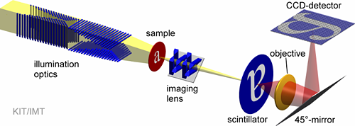
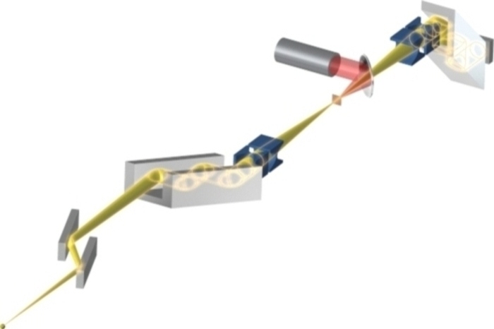
Schematic view of an X-ray full-field microscope
 |
 |
 |
||
| Parabolic line focus lens | Point focus lens | X-ray prism lens |
Read more…
 |
 |
|
 |
 |
|
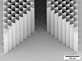
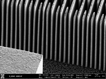
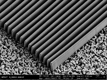
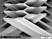
| 1 µm period gratings (left: “bridge design” absorption gratings; gold thickness: 20 µm; right: long lamellae phase gratings; nickel thickness: 3 µm) |
||
 |
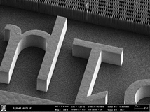
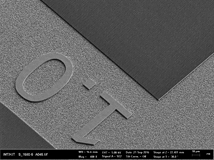
| X-ray LIGA KIT logo in gold in front of a 10 µm period 300 µm high grating with sun-rays (before electroplating) (courtesy of JulesMarketing). |
We continuously improve the gratings quality with focus on the following issues:
- repeatability of the process,
- downsizing the period and increasing the resist/metal thickness by modifying the photoresist sensitivity and contrast,
- increasing the mechanical stability by finding the best combination of applied dose and post exposure bake (PEB), by using freeze drying and room temperature electroplating,
- minimizing the harmful effects of secondary radiation by using a low Z electroplating layer/substrate, optimizing the design and the gratings’ geometry to obtain the minimum possible deviation from an ideal binary grating,
The characterization is done using:
- scanning electron microscopy (DC measurement),
- white light interferometry (G1 measurement thickness)
- radiography, visibility measurement using standard grating setups by our partners (CT-Lab at KARA, TUM, FAU).
X-ray lenses made out of polymer by KIT/IMT have been compared to beryllium, carbon and diamond lenses at beamline Petra III-P01: they are the only ones free of birefringence and dichroism and thus the only lenses suitable for polarization experiments.

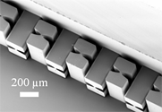
The recently developed staircase array of inclined refractive multi-lenses can perform large field of view (FoV) pixel super-resolution scanning transmission hard X-ray microscopy with a 780 ± 40 nm spatial resolution within a FoV of 1.64 cm × 1.64 cm. (Photo: IUCr Journals, Wiley)
DOI: 10.1107/S1600577521001521
More…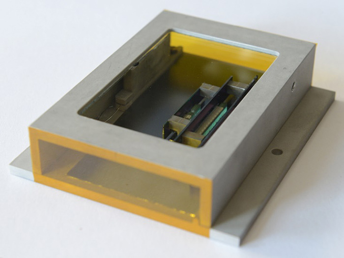
Compound refractive X-ray lenses (CRLs) made by KIT/IMT have been used to upgrade the high-pressure X-ray diffraction (XRD) beamline BL10XU at the synchrotron source SPring-8 in Japan to enable realizing diffraction experiments of micrometer-sized samples at multi-megabar pressures (Photo: Arndt Last, KIT).
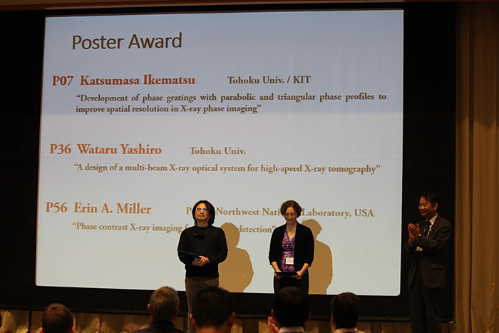
Dr. Katsumasa Ikematsu (KIT and Tohoku University, Sendai, Japan) received the Poster Award of the 5th XNPIG in Sendai (Japan) for his contribution on the improvement of the spatial resolution in X-ray phase imaging (Photo: LOC XNPIG2019).

The foundation supports only about 8% of the applications and supports the innovative proposal even without the usual oral presentation. A liquid X-ray lens could be used in the white X-ray beam of a synchrotron source or a free-electron laser, since heating and damage to the lens are constantly cured.
Liquid X-ray lens
Two of the three collaborative research projects which are funded by the BMBF on the topic of “Optics and Photonics” are headed by IMT. Next to Japanese partners from academia and industries the spin-off from KIT, microworks and Vanguard Photonics, are also part of the consortia (Photo: BMBF).
The block course X-ray optics, which starts at June 4th, will not only teach you in the fundamentals of X-ray optics and X-ray imaging but will also give information on X-ray imaging in medical diagnostic and introduce new methods in radiography and computed tomography like phase contrast and dark field imaging.
More…
In the last 10 years, from 2008 to 2017, at the synchrotron source SPring-8 in Japan 225 publications have been published where compound refractive X-ray lenses (CRLs) from KIT/IMT have been used in the experimental setup. This makes a total of nearly 1500 pages published by scientists at the beamlines BL10XU, BL13XU, BL15XU, BL22XU, BL24XU, BL41XU and BL43LXU. All these papers are based on measurements where CRLs from IMT helped illuminating the sample in diverse experimental methods like for example micro diffraction, resonant scattering, micro-XAFS, GISAXS or ultra-high pressure research.
ESRF announced that the Full Field Diffraction X-ray Microscopy (FFDXM) end station at the ID01 is now open to user experiments. This new method uses X-ray lenses from KIT/IMT specially designed for the imaging part of the setup.