

X-ray Micro-Technology (XMT)
Prof. Dr. Venera Weinhardt [Contact]
Welcome
...to the Weinhardt lab! X-ray imaging is a powerful technology that has been extensively used in medical diagnosis and industrial non-destructive inspection. We are working in this exciting, fast-developing field, with expertise at the interface of micro- and nanofabrication, optical technologies, and machine learning.
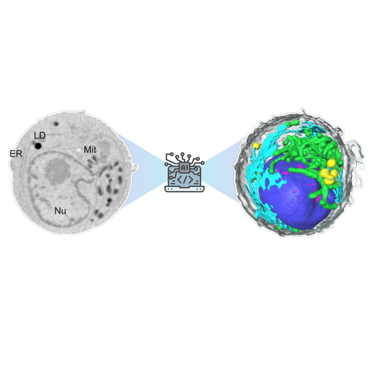
Our department works on technological developments in micro- and nano-X-ray imaging across multiple structural scales, focusing on laboratory solutions accessible to the broader research community. We develop novel sample preparation technologies, imaging methods, and algorithms for quantitative analysis, including AI-driven image segmentation methods. For our work, we have established a set of tools and resources available only within our department. Our research contributes to the research program 3: "Materials Systems Engineering" (MSE), Topic 5: "Materials Information Discovery", Link to MSE website.
Research Groups
X-ray Imaging
.png)

We are happy to announce that X-ray optics developed at IMT could help decipher dynamic strain effects in oxide thin-film systems. In a new Science publication, researchers show that substrates are not only passive supports, but can actively participate in the structural response of thin-film materials.
Dr. Arndt Last from the Weinhardt Lab contributed his expertise in polymer compound refractive X-ray lenses, supporting the high-resolution X-ray imaging approach used in the study.
More
From 08 to 10 July 2026, the X-ray Micro-Technologies Department at IMT will host the workshop “Correlative Imaging, Information Theory, and Characterization” at KIT Campus North.
The program includes scientific talks and hands-on sessions with imaging demonstrations, data analysis activities, and sample characterization.
We warmly invite interested participants to join the workshop.
Further Information and Registration
It was a great pleasure to welcome 24 curious and creative young girls to our department at IMT as part of this year’s Girls’ Day. We were showcasing the fascinating world of X-ray imaging, where girls could transform science into art using simple everyday objects. X-rays can be literally insightful! A big thank you to my team and to 3DMM2O for making this event so inspiring. We’re excited to see the next generation of scientists take their first steps into research.
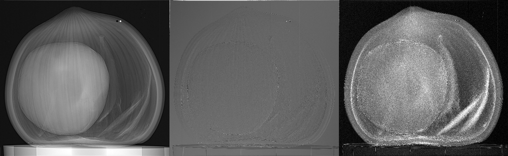
IMT researchers at KIT published "SHI: a framework for spatial harmonic imaging" in Scientific Reports. SHI is a modular Python framework for multicontrast X-ray imaging using periodic modulators (e.g., Hartmann masks). It automates acquisition, higher-order-harmonic retrieval, and CT preprocessing, extracting absorption, refraction, and scattering signals from a single exposure. Robust to large focal spots and optical imperfections, it reveals internal structures across a range of contrasts with fewer projections, reducing dose.
Read the full manuscript here