

Multimodal X-ray Imaging (MXI)
Prof. Dr. Venera Weinhardt [Contact]
Welcome
The MXI Lab develops innovative imaging techniques at the macro- and nanoscale across a range of biological scales, emphasizing user-friendly laboratory solutions to make these advanced tools accessible to the biomedical community.
Subject areas
We develop X-ray imaging methods for different hierarchical scales.
Correlative light and X-ray microscopy
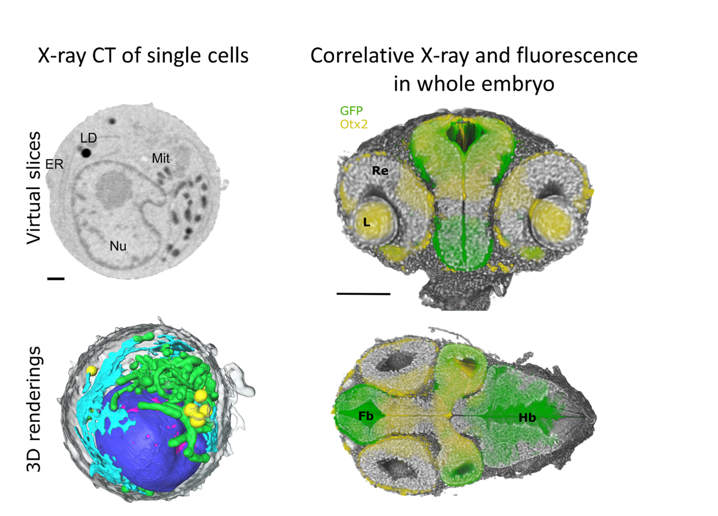
Morphometric data on cells are of limited use without knowledge of their functions. Physiological processes such as movement, ion transport, and metabolism, including protein location and associations, are typically visualized by fluorescent light microscopes. Therefore, we are working on the correlation of light and X-ray microscopy. Within the MSCA-DN CLEXM project [link], we integrate full-rotation soft X-ray tomography (SXT)—established for isotropic 3D cellular ultrastructure—with axial super-resolution fluorescence microscopy adapted for thin glass-wall capillaries. Full-rotation tomography uniquely delivers isotropic nanometer-scale 3D data, combining SXT's label-free, quantitative density mapping (e.g., distinguishing hetero- from euchromatin via X-ray absorption) with fluorescence for molecular specificity, to probe dynamic chromatin reorganization in biological processes.
Automatic high-throughput X-ray tomography
Within the “SMART-Morph” project [link], we develop a fully automated pipeline for high-throughput sample preparation for X-ray imaging, leveraging optical microscopy integrated with custom AI algorithms to enable rapid selection, sorting, labeling, and grouping of samples (e.g., by developmental stage) into labeled holders and trays. These automatic sample preparation systems directly feed into robotic X-ray sample changers for seamless beamline delivery, minimizing manual intervention and scaling workflows for synchrotron or lab-based tomography. This addresses key bottlenecks in X-ray microscopy by ensuring consistent positioning, traceability, and batch processing, boosting throughput for 3D cellular and tissue analysis in biomedical research.