

Magic Angle Coil Spinning (MACS) Detectors
(Dr. Vlad Badilita, Prof. Jan G. Korvink, Shyamsunder Adhikari)
At SPA Lab, we employ MEMS fabrication techniques to develop advanced miniaturised detectors for nuclear magnetic resonance (NMR) spectroscopy of sub-microliter samples. The so-called MACS detectors are being used in the magic angle spinning (MAS) configuration, the main difference being that here, both the detector and the sample rotate rapidly in the magnetic field and the signal is transmitted via inductive coupling between the MACS coil and the static coil (Figure 1).
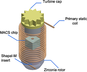
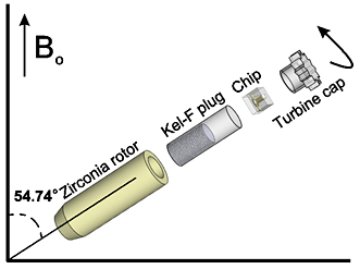
Figure 1: a) Schematic of the MACS detector inserted in the primary coil; b) Exploded view of the Magic Angle Coil Spinning arrangement.
The MACS detector (Figure 2a) consists of a wirebonded solenoidal microcoil (the inductance - L) and an on-chip capacitor (the capacitance - C). The values for L and C can be independently chosen in order to achieve the Larmor frequency of interest. This microcoil-capacitor design leads to a versatile sensor able to cover a wide range of Larmor frequencies. The fabrication is based on a wafer level process providing a high degree of reproducibility, robustness, as well as a small end-price per detector.
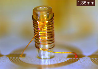
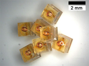
Figure 2: (a) MACS detector before encapsulation in SU-8 showing the high aspect ratio wirebonded microcoil and the on-chip interdigitated capacitor; (b) Encapsulated MACS detectors as disposable, single-use devices.
We anticipate that the MACS detectors will become consumable, single-use detectors (Figure 2b) used in a wide area of applications ranging from metabolomics to solid state NMR characterisation. Figure 3a shows an NMR spectrum from an early stage Drosophila pupae where, after suppressing the side-bands due to spinning, as well as the dominant water peak, the individual metabolites within the sample can be identified. Figure 3b shows the prospect of using the MACS detectors for solid state applications, demonstrating line-width narrowing of adamantane with increasing the spinning speed. It is important to note that the volume of each sample is about 330 nl.
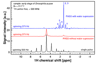
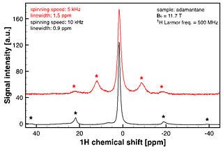
Figure 3: (a) Metabolomics applications of MACS detectors: identifying metabolites in Drosophila pupae (graph and full info); (b) Solid state NMR applications of MACS detectors: line-width narrowing with increased spinning speed for an adamantane sample (from https://dx.doi.org/10.1371/journal.pone.0042848).Each sample has a volume of 330 nl.
Furthermore, Figure 4 illustrates the use of the MACS insert as a tool for high-resolution single embryo metabolomic profiling using 100 hpf (hours post fertilization) zebrafish embryos: proton and 1H-1H COSY MAS NMR spectra.
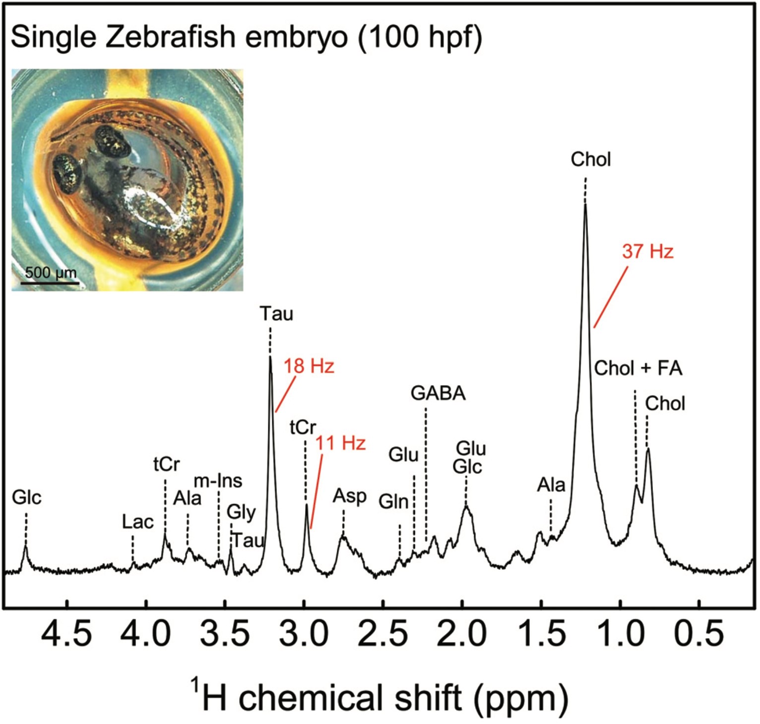
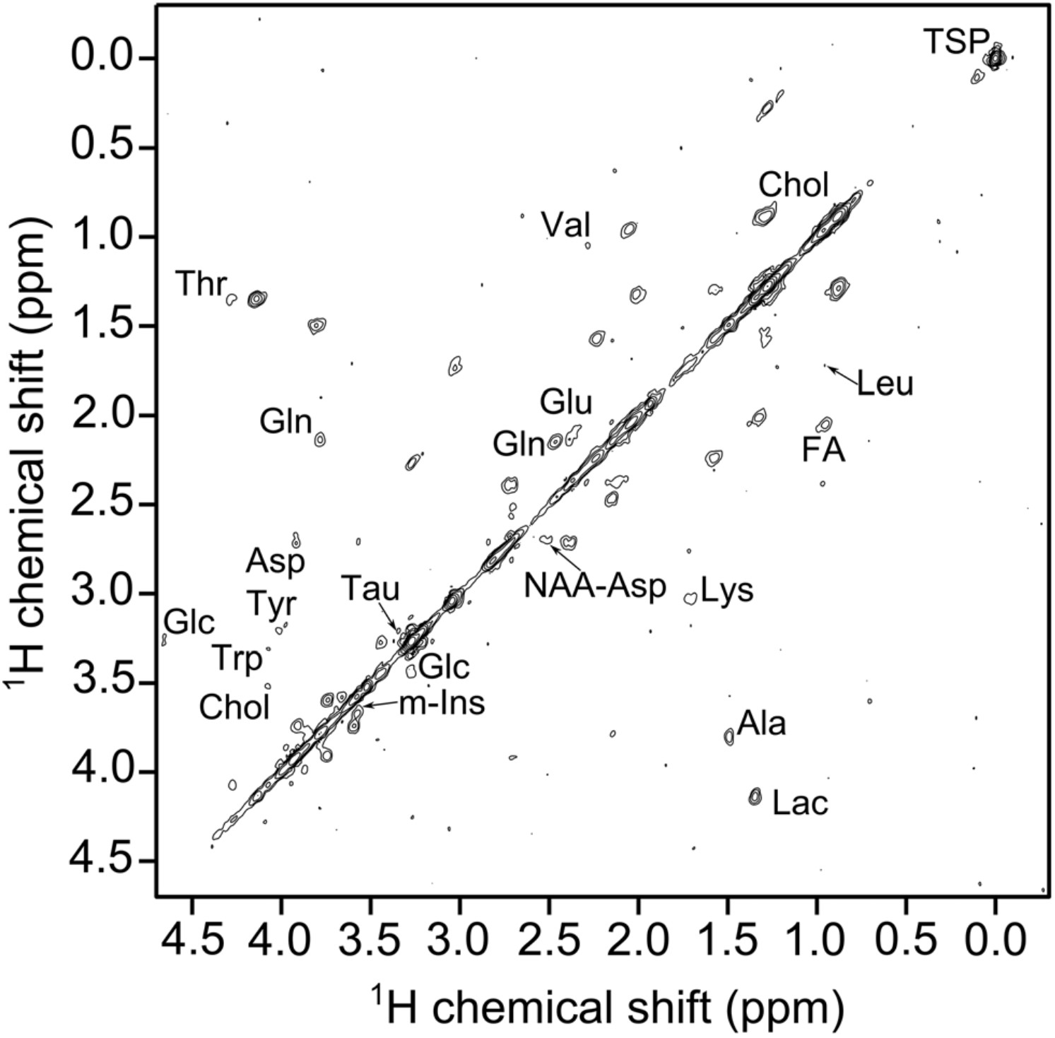
Figure 4: (a) 1H MAS NMR spectrum of a single zebrafish embryo spun at 5 kHz after solvent suppression. The metabolic profile of the embryo shows a number of fatty acids, sugars etc. among other metabolites (Ala, alanine; Asp, aspartate; Glc, glucose; Gln, glutamine; Glu, glutamate; Gly, glycine; Lac, lactate; Tau, taurine; m-Ins, (myo)-inositol, tCr, total creatine; GABA, gamma-aminobutyric acid; Chol, cholesterol; FA, fatty acid) The inset shows the photograph of an intact zebrafish embryo on top of the MACS orifice before loading; (b) 1H–1H COSY NMR spectrum of a single zebrafish embryo under MAS condition of 5 kHz after solvent suppression using a 20 μW pulse. Additional metabolites like tyrosine, leusine, L-tryptophan (Trp) etc. are detected. (graph and full info)
MACS are just one type of detector from our NMR detector portofolio. Please check out our other NMR sensors, e.g., Helmholtz coils (more information), broadband spiral coils , stripline Lenz lenses (DOI: 10.1038/s41598-023-50616-0).